Nanomedicine | BioMEMS/NEMS Biosensors/Biomolecule Capture and Sorting | Single Molecule Imaging | Virology | In Silico Biology
Microbotics: Bacteria-mediated delivery of smart nanocargo into cells
Akin,
D., J. Sturgis, K. Ragheb, D. Sherman, K. Burkholder, J. P.
Robinson, A. K. Bhunia, S. Mohammed and R. Bashir.
Bacteria-mediated delivery of nanoparticles and cargo into cancer
cells. Nature Nanotechnology, 2:441-449, 2007. (download PDF)
Nanoparticles and bacteria have been independently used to deliver
genes and proteins into mammalian cells for monitoring or altering gene
expression and protein production. Here, we show the simultaneous use
of nanoparticles and bacteria to deliver nucleic acid-based model drug
molecules into cells and mice. In our approach, the gene or cargo is
loaded onto the nanoparticles, which are carried on the bacteria
surface. The bacteria successfully delivered the molecules, and the
genes were released from the nanoparticles and expressed in four
different cell types and mice. This new approach may be used to deliver
different types of cargo into a variety of cells and live animals
without the need for complicated genetic manipulations.
Bioinspired-Cancer Drug Delivery "Cellular Trojan Horses"
Choi,
M.R., Stanton-Maxey, K.J., Stanley, J.K., Levin, C.S., Bardhan, R.,
Akin, D., Badve, S., Sturgis, J., Robinson, J.P., Bashir, R., Halas,
N.J., Clare, S.E. A Cellular Trojan Horse for Delivery of
Therapeutic Nanoparticles into Tumors. Nano Letters, In Press, 10.1021/nl072209h S1530-6984(07)02209-6, 2007.
Destruction of hypoxic regions within tumors, virtually inaccessible to
cancer therapies, may well prevent malignant progression. The tumor's
recruitment of monocytes into these regions may be exploited for
nanoparticle-based delivery. Monocytes containing therapeutic
nanoparticles could serve as "Trojan Horses" for nanoparticle transport
into these tumor regions. Here we report the demonstration of several
key steps toward this therapeutic strategy: phagocytosis of Au
nanoshells, and photoinduced cell death of monocytes/macrophages as
isolates and within tumor spheroids.
Solid-state Nanopore Channels with DNA Selectivity
Solid-state nanopores have emerged as possible candidates for
next-generation DNA sequencing devices. In such a device, the DNA
sequence would be determined by measuring how the forces on the DNA
molecules, and also the ion currents through the nanopore, change as
the molecules pass through the nanopore. Unlike their biological
counterparts, solid-state nanopores have the advantage that they can
withstand a wide range of analyte solutions and environments. Here we
report solid-state nanopore channels that are selective towards single
strand DNA (ssDNA). Nanopores functionalized with a 'probe' of hair-pin
loop DNA can, under an applied electrical field, selectively transport
short lengths of 'target' ssDNA that are complementary to the probe.
Even a single base mismatch between the probe and the target results in
longer translocation pulses and a significantly reduced number of
translocation events. Our single molecule measurements allow us to
separately measure the molecular flux and the pulse duration, providing
a tool to gain fundamental insight into the channel-molecule
interactions. The results can be explained in the conceptual framework
of diffusive molecular transport with particle-channel interactions.
Biomedically Relevant Nanomaterials and their Biocompatibility
Bajaj,
P.,D. Akin, A. Gupta, O. Auciello and R.Bashir.
Ultrananocrystalline diamond film as an optimal cell interface for
biomedical applications.Biomedical Microdevices, I9:787-94, 2007.
Surfaces of materials that promote cell adhesion, proliferation, and
growth are critical for new generation of implantable biomedical
devices. These films should be able to coat complex geometrical shapes
very conformally, with smooth surfaces to produce hermetic bioinert
protective coatings, or to provide surfaces for cell grafting through
appropriate functionalization. Upon performing a survey of desirable
properties such as chemical inertness, low friction coefficient, high
wear resistance, and a high Young’s modulus, diamond films emerge as
very attractive candidates for coatings for biomedical devices. A
promising novel material is ultrananocrystalline diamond (UNCD®) in
thin film form, since UNCD possesses the desirable properties of
diamond and can be deposited as a very smooth, conformal coating using
chemical vapor deposition. In this paper, we compared cell adhesion,
proliferation, and growth on UNCD films, silicon, and platinum films
substrates using different cell lines. Our results showed that UNCD
films exhibited superior characteristics including cell number, total
cell area, and cell spreading. The results could be attributed to the
nanostructured nature or a combination of nanostructure/surface
chemistry of UNCD, which provides a high surface energy, hence
promoting adhesion between the receptors on the cell surface and the
UNCD films.
Micro/Nanoscale cantilevers as biosensors
Gupta, A., P.R. Nair,D. Akin, M, Ladisch, S. Broyles, M. A. Alam and R.
Bashir. Anomalous resonance in a nanomechanical bioSensor. Proc. Natl. Acad. Sci., USA, 103:13362-13367, 2006.
Normally a cantilever's resonant frequency decreases when molecules
attach to it – a finding that is the basis of nanomechanical sensing
devices- but we have found that the resonant frequency of some
nanoscale cantilevers may actually increase on the addition of
molecules. Area-dependent protein adsorption is shown in the side
figure. (a) Schematic diagram depicting the methodology of the specific
binding of the secondary Abs to the proteins used in scheme 1. (b)
Photomicrograph of fluorescently labeled (FITC; green) Ab to BSA
attached to varying sized cantilever beams clearly showing an increase
in fluorescent intensity for longer cantilevers. (Scale bar, 5 μm.) (c)
Semilog plot showing the measured average fluorescence intensity from
the secondary Abs to the proteins used in scheme 1 as a function of
cantilever beam area. (Inset) The same parameters in the linear scale.
The squares indicate the simulated protein density at the tip of the
cantilevers shown in b. The simulated value of the shortest cantilever
beam (LC = 5 μm in b) was normalized with the measured value of the
same length scale, the remaining two simulated lengths (LC = 10 and 15
μm) were scaled by the same factor, and then all three simulated values
were plotted with the measured data. (d) Simulated protein density
distribution on the adsorbing cantilever surfaces. The density reaches
a maximum for the longer cantilever. The monotonic increase in density
with the cantilever length is due to the competitive attachment of
protein among the adsorbing surfaces. Simulated protein density at the
tip of the cantilevers is in excellent agreement with the experimental
results.
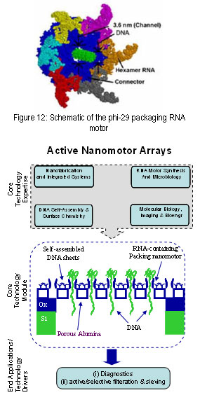
Biohybrid Nanodevices for Nanomedicine
Use of Bacteriophage Phi-29 Packaging RNA NanoMotor for Active Devices for Nanomedicine:
Demir Akin, Peixuan Guo, Chengde Mao, and Rashid Bashir,
A specific project funded through NIH Nanomedicine Center involves the
use of the Phi-29 packagaing RNA nanomotor and interfacing this
biological motor with micro/nano fabricated devices. The center
overview can be found at (http://www.vet.purdue.edu/PeixuanGuo/NDC/).
The goal of the proposed Nanomedicine Development Center (NDC):
“Phi29 DNA-Packaging Motor for Nanomedicine”, is to create biologically
compatible membranes and arrays with embedded and active phi29
DNA-packaging motors for applications in medicine. For example,
currently there is no nanodevice available for actively pumping drugs,
DNA/RNA and other therapeutic molecules into specifically targeted
cells. Our NDC, (also referred to as the Nanomotor Drug Development
Center, NDDC), will create a hybrid system that combines the best
features of the biological motor with synthetic delivery systems that
have already achieved clinical success. The re-engineered motors
developed will also be applied in various array formats to extend
application to diagnostics and other therapeutic approaches. One of the
thrusts is to develop novel diagnostic and therapeutic devices by
integrating the phi-29 motor to micro/nano fabricated surfaces. We are
working on making arrays of these motors for possible application
selective filteration and sieving devices. Specifically we are working
on use of surface fucntionalization techniques to form motor arrays on
silicon surfaces and demonstrate the operation of motor arrays. Next we
will work on use of nanoporous membranes and attempt to attach the
nanomotors on these membranes in hopes to make selective sieving and
filteration devices.
Normally
a cantilever's resonant frequency decreases when molecules attach to it
– a finding that is the basis of nanomechanical sensing devices- but we
have found that the resonant frequency of some nanoscale cantilevers
may actually increase on the addition of molecules.
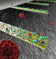
Shown here is an array of functionalized cantilvers. An array of tiny,
diving-boardlike devices called nanocantilevers. The devices are coated
with antibodies to capture viruses, which are represented as red
spheres. New findings about the behaviour of the cantilevers could be
crucial in designing a new class of ultra-small sensors for detecting
viruses, bacteria and other pathogens. (Image generated by Seyet, LLC).
A. Gupta, P. Nair, D. Akin, S. Broyles, M. Ladisch, A. Alam, R. Bashir,
"Anamolous Resonance in a Nanomechanical Biosensor", Proceedings of
National Academy of Sciences, USA. August 28, 2006,
10.1073/pnas.0602022103 - (download PDF)
Cell mass sensing and measurement of growth changes:
Park, K., J.Jang,D. Akin, D. Irimia, M. Toner, and R. Bashir. Capture,
growth and mass measurement of mammalian cells on silicon cantilever
arrays. Biomedical Engineering Society, September 22, 2007, Los
Angeles, LA.
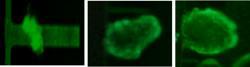
Tissue Engineering and Biohybrid Devices
Our
improved understanding of how biological systems, from proteins to
subcellular compartments to cells, to tissues, to organs, and
eventually, to the entire organism are formed and regulated, and the
nanoscale control of sythetic material physicocehmical properties will
enable us to devise and realize the next generation of nanomedical
systems for improvement of human health. Towards these goals, we adopt
the emerging cutting edge biomedical research findings into engineering
and perform research and developement in biohybrid devices. One example
of these is given in the figure in the right column. A microfabricated
cantilver is surface functionalized and embryonic cardiomyocytes are
grown on it, forming a beating sheet of cardiac tissue that actuates
the cantilever. These types of devices have desirable properties for
numerous areas of bioinspired and engineered biomedical solutions, from
drug screening to bidirectional signal conversion between biological
and electronic signals, bioenergy to drug delivery, to artificial
organs.