1996 Project Reports | Home |
Contents | Previous |
Next |
Bone remodeling simulations - validation using quantitative computed
tomography
Gary Beaupré, PhD; Greg Breit, PhD; Robert Whalen, PhD; Dennis
Carter, PhD; John Drace, MD; Virginia Giddings, MS; Jenny Kiratli, PhD; Sandy
Napel, PhD; Inder Perkash, MD; George Sims, MD; Chye Hwang Yan, MS
Our understanding of how bone adapts in response to mechanical stimuli has
expanded considerably in the past ten years in large part because of the
development of new mathematical-based theories for bone remodeling. Some of
these theories have undergone preliminary validation through computer
simulations of bone development, adaptation from altered loading, and bone
remodeling after prosthetic implantation. While the majority of these
validation studies have been qualitative in nature, they have suggested new
insights into the mechanical factors that influence skeletal biology. In spite
of these advances there is a compelling need for additional, quantitative
validation studies.
With the help of collaborators in the Departments of Radiology, Spinal Cord
Injury and Orthopaedics we have begun a study that will use data obtained from
VA patients to validate and refine the bone remodeling theory developed at the
VA Rehabilitation R&D Center and Stanford University. The patients
participating in this study have sustained either a spinal cord injury or an
ankle fractures. Both of these patient groups experience significant bone loss
(osteoporosis) in the heel bone of the foot. We are measuring the amount of
bone loss in these patients since their injury, using a very precise technique
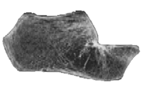
called Quantitative Computed Tomographym or QCT (Fig. 1). Using QCT we can
monitor not only the total amount of bone loss but also the distribution of
bone loss throughout the internal volume of the heel bone. By comparing the
measured bone loss with the predictions from computer models we can validate
and refine our bone remodeling algorithm with an accuracy and precision never
previously achieved.
 Figure
1. CT scan of the calcaneus (heel bone).
Another important question that we are examining in this study concerns the
extent to which bone loss is reversible. Our QCT measurement in patients with
ankle fractures should help to answer this question. Some of the bone that was
lost during the cast immobilization phase should be recovered after cast
removal. However, it is possible that complete recovery of bone mass will not
occur in some patients. While the reasons for this are not entirely clear, the
ability to monitor bone mass in patients using QCT will allow us to examine the
role of mechanical forces in the process of bone recovery and possibly suggest
new rehabilitation strategies.
|
Republished from the 1996 Rehabilitation R&D Center Progress Report. For
current information about this project, contact:
Gary Beaupré.

