1996 Project Reports | Home |
Contents | Previous |
Next |
Tissue differentiation - understanding the implant/bone interface
Gary Beaupré, PhD; Nick Giori, PhD; MD Dennis Carter, PhD; Stuart
Goodman, MD
One of the most challenging problems limiting the long-term success of hip
and knee replacements is implant loosening. A loose and painful implant
typically requires revision surgery for removal of the prosthesis and
reimplantation with a new prosthesis. For more than 30 years, bone cement has
been used to secure the implant to the supporting bone. However, the
unavoidable accumulation of fatigue cracks in the cement inevitably leads to
implant loosening and the need for revision.
During the 1980's researchers and implant manufactures developed a new
generation of implants that no longer relied on the use of cement for implant
fixation. With these cementless designs it was thought that fixation would
occur via the generation or differentiation of new bone between the implant and
the adjacent bony bed (Fig. 1). The process of bony fixation, however, has
proven to be both unpredictable and unreliable. Frequently, a layer of fibrous
tissue rather than bone develops at the implant-bone interface.
 Figure
1. Close-up of the implant-bone interface surrounding a joint replacement.
We are working on the development of theoretical models (Fig. 2) that will
enable us to predict the type of interface tissue that will develop or
differentiate around an implant in response to the local stress and strain
environment. Using a novel implantable chamber made out of pure titanium (Fig.
3) we are studying the influence of shear strain on tissue differentiation in
an animal model. These types of studies are essential if we are to understand
the mechano-biological processes controlling the biological fixation of
cementless implants. This knowledge, in turn, will lead to the development of
new implant designs that will function better and last longer than current
joint replacements.
|
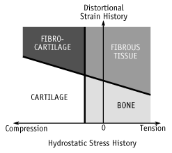 Figure 2. Palo
Alto VA/Stanford tissue differentiation hypothesis.
|
 Figure 3.
Exploded view of titanium tissue differentiation chamber.
|
Republished from the 1996 Rehabilitation R&D Center Progress Report. For
current information about this project, contact:
Gary Beaupré.
