Easy-DHPSF V2.0: open-source software for three-dimensional localization and two-color registration of single molecules with nanoscale accuracy
Version 2.0 (April 2019)
For download capability, see http://sourceforge.net/projects/easy-dhpsf
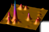
Automated processing of double-helix (DH) microscope images of fluorescent single molecules (SMs) streamlines the protocol required to obtain super-resolved three-dimensional (3D) reconstructions of ultrastructures in biological samples by single-molecule active control microscopy (SMACM). Here, we present a suite of MATLAB subroutines, bundled with an easy-to-use graphical user interface (GUI), that facilitates 3D localization of single emitters (e.g. SMs, fluorescent beads, or quantum dots) with typical precisions of tens of nanometers in multi-frame movies acquired using a wide-field DH epifluorescence microscope. The algorithmic approach is based upon template matching for SM recognition and least‑squares fitting for 3D position measurement, both of which are computationally expedient and precise. While this approach is straightforward, overlapping images of SMs are ignored, and the precision of least-squares fitting is not as high as maximum likelihood-based methods. Version 2.0 of Easy-DHPSF augments the approach of Version 1.0 by incorporating code that facilitates combining localization data from two spectral channels using a locally-weighted quadratic 3D registration function.
Camille Bayas, Alex von Diezmann, Anna-Karin Gustavsson, and W. E. Moerner, "Easy-DHPSF 2.0: open-source software for three-dimensional localization and two-color registration of single molecules with nanoscale accuracy," 24 April 2019, PROTOCOL (Version 2) description available at Protocol Exchange [https://doi.org/10.21203/rs.2.9151/v2]. pdf
Code available at sourceforge.net/projects/easy-dhpsf
____________________________________________________________________
Easy-DHPSF open-source software for three-dimensional localization of single molecules with precision beyond the optical diffraction limit
Version 1 (2013)
For download capability, see http://sourceforge.net/projects/easy-dhpsf
Automated processing of double-helix (DH) microscope images of single molecules (SMs) streamlines the protocol required to obtain super-resolved three-dimensional (3D) reconstructions of ultrastructures in biological samples by single-molecule active control microscopy. Here, we present a suite of MATLAB subroutines, bundled with an easy-to-use graphical user interface (GUI), that facilitates 3D localization of single emitters (e.g. SMs, fluorescent beads, or quantum dots) with precisions of tens of nanometers in multi-frame movies acquired using a wide-field DH epifluorescence microscope. The algorithmic approach is based upon template matching for SM recognition and least-squares fitting for 3D position measurement, both of which are computationally expedient and precise. Overlapping images of SMs are ignored, and the precision of least-squares fitting is not as high as maximum likelihood-based methods. However, once calibrated, the algorithm can fit 15-30 molecules per second on a 3 GHz Intel Core 2 Duo workstation, thereby producing a 3D super-resolution reconstruction of 100,000 molecules over a 20×20×2 μm field of view (processing 128×128 pixels × 20000 frames) in 75 min.
Matthew D. Lew*, Alexander R. S. von Diezmann*, and W. E. Moerner, Protocol Exchange, doi:10.1038/protex.2013.026, published online 25 February 2013. DOI[ Slide] pdf Slide] pdf
|