

Pulsed Electron Avalanche Knife (PEAK)
Electrosurgery, one of the most common surgical technologies, is considered a robust but somewhat crude technique, with its basics changed surprisingly little since its invention almost a century ago. Continuous radiofrequency is still used for tissue cutting, with thermal damage extending to hundreds of micrometers. In contrast, lasers developed seventy years later, have been constantly perfected, and the laser-tissue interactions explored in great detail, which has allowed tissue ablation with cellular precision in many laser applications.
Pulsed electrosurgery with properly optimized waveforms and microelectrodes can rival many advanced lasers. Similar to lasers, spatial and temporal confinement of energy deposition in electrosurgery can greatly improve precision and reduce collateral damage. Pulsed plasma-mediated discharges with burst durations in tens of microseconds applied via insulated planar electrodes with micrometers-wide exposed edges can dissect tissues with the collateral damage zone not exceeding cellular scale. Length of the electrodes can vary from micrometers to centimeters, and all types of soft tissues – from membranes to cartilage and skin could be dissected in liquid medium and in a dry field.
Interactions of pulsed electric field with biological tissues include heat generation and diffusion, coagulation, vaporization, cavitation, ionization, as well as non-thermal interactions such as electroporation and neural stimulation. We designed electrode configurations and pulsed waveforms for minimally-traumatic surgical and therapeutic applications.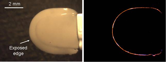
Left: A blade electrode with insulated sides and exposed narrow edge.
Right: Plasma discharge in physiological medium along the exposed metal edge
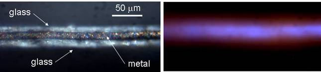
Left: Higher magnification side view of the blade demonstrates 12 μm metal foil and 10 μm layers of glass insulation.
Right: Light emission from the plasma discharge along the edge of the blade.
Waveforms of electric current and light emission by the plasma during the burst. Phases of initial heating, vaporization and ionization are shown by the arrows.
Cut in the human retina produced by PEAK in-vitro.
SEM micrograph of the rabbit lens capsule cut with PEAK. Note the sharpness of the edge - the scale bar is 10 μm.
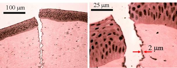
Cut in a porcine cornea produced by PEAK in-vitro. Note the 2 μm width of the thermal damage zone (darker area at the edges of the lesion).
We called this technology Pulsed Electron Avalanche Knife (PEAK). It was commercialized by PEAK Surgical Inc., and is currently manufactured by Medtronic, with world-wide distribution for a wide variety of surgical applications (PEAK Plasmablade).