

Coherent Anti-Stokes Raman Scattering (CARS) Microscopy
The low natural optical contrast of biological cells and tissues necessitates staining of such samples with various chromophores and fluorescent markers. However, exogenous staining often affects cellular metabolism and thus is of limited utility for imaging of living cells. CARS microscopy allows for imaging living cells with intrinsic chemical contrast. Similarly to spontaneous Raman scattering, CARS is sensitive to the molecular structure of the material and its efficiency is greatly enhanced in the vicinity of the molecular vibrational resonances. The coherent nature of this process can make the CARS signal much greater than that of spontaneous Raman scattering.
Since the first demonstration of CARS microscopy in 1982, and through its extensive development during the last decade, practically all methods have been based on scanning of tightly focused co-propagating pump and Stokes laser beams.
We developed a wide-field illumination scheme for picosecond CARS microscopy, in which the whole image is registered simultaneously - similar to conventional microscopy with a focal plane array detector. Such microscopes could have faster image acquisition rates, significantly relaxed requirements on laser stability, and simpler mechanical design with none of the moving parts inherent to scanning systems.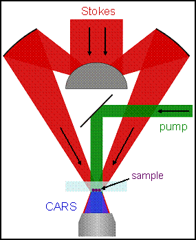
To prevent CARS generation in a homogeneous bulk material, we use a non-phase-matching illumination, and rely on light refraction or scattering within the sample that brings some portion of the incident radiation into the phase matching condition. Thus, the CARS signal is generated only in the presence of a scattering or refracting object. If such an object, or its immediate surrounding, contains molecules with Raman-active vibrational modes, the CARS signal will be greatly enhanced. In this respect, such illumination geometry resembles dark field illumination in conventional microscopy. However, nonlinear optical transformation confers to this technique a new quality – the CARS signal, which depends on both the optical properties of the object and its chemical composition.
Wide-field illumination geometry for inverted microscope. Stokes beam is focused by a reflective (Cassegrain) objective, while the collimated pump beam is reflected onto the sample from a flat mirror. CARS image is acquired via a conventional microscope objective located below the sample.
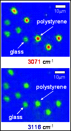
Spectral selectivity of CARS: Polystyrene beads imaged at 3071 cm-1 resonance (top) exhibit much stronger signal than out of resonance at 3116cm-1 (bottom). For comparison, sample included glass beads of similar size (6 μm), which have no Raman resonance at these wavelengths.
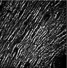 Selective imaging of the lipid-rich myelin sheaths in the spinal cord tissue, using aliphatic C-H stretch resonance at 2914 cm-1.
CARS microscope can be used for selective imaging of specific structures in living cells and tissues without exogenous staining, and for monitoring dynamics of cellular changes under conditions of physiological stress.