dystrophin in dysferlin-deficient
              A/J skeletal muscle  
Stanford University  
line decor
    HOME  ::  
line decor


Research Interests
 

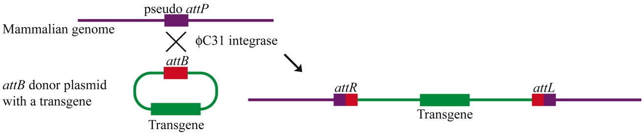
The Calos Lab is interested in developing novel gene and cell therapy approaches to address human diseases.

Our primary approach is to use genetically-corrected stem cells as therapeutics, to develop strategies to treat genetic diseases like muscular dystrophy.

The lab has expertise in methods for the genetic engineering of mammalian cells, including site-specific integrases, homologous recombination, and extrachromosomal vectors.


Phage integrases for recombination in mammalian cells

For example, we have developed the phiC31 integrase system for use in mammalian cells. This phage integrase pairs two short recognition sites, called attB and attP, and catalyzes recombination that results in chromosomal integration.

This figure shows one approach we have developed with phiC31 integrase. We place an attB site on the plasmid that we wish to integrate into the chromosome. The enzyme finds a site similar to attP in the chromosome and carries out recombination. The net result is integration of the plasmid into the chromosome:


integration reaction

  • Groth AC, Olivares EC, Thyagarajan B, Calos MP. (2000). A phage integrase directs efficient site-specific integration in human cells. Proc Natl Acad Sci U S A. 97(11):5995-6000. [Abstract]
  • Thyagarajan B, Olivares EC, Hollis RP, Ginsburg DS, Calos MP. (2001). Site-specific genomic integration in mammalian cells mediated by phage φC31 integrase. Mol Cell Biol. 21(12):3926-34. [Abstract]

We also utilize integrases with higher specificity, like Bxb1, that can recombine only at their own att sites. If the Bxb1 attP site is placed into the chromosomes, then Bxb1 integrase can be used to recombine a plasmid bearing the Bxb1 attB site into this location with high specificity. This short movie created by Alfonso Farruggio, a Genetics graduate student in the lab, illustrates how the reaction works:


  • Chalberg TW, Portlock JL, Olivares EC, Thyagarajan B, Kirby PJ, Hillman RT, Hoelters J, Calos MP. (2006). Integration specificity of phage φC31 integrase in the human genome. J Mol Biol. 357(1):28-48. [Abstract]


Applications in Gene Therapy


We have developed approaches that cure hemophilia in disease model mice, by using hydrodynamic DNA injection and phiC31 integrase to bring about permanent integration of the factor VIII or IX genes in the liver:

  • Keravala, A., Chavez, C.L., Hu, G., Woodard, L.E., Monahan, P.E., and Calos, M.P. (2011) Long-term phenotypic correction in factor IX knockout mice by using phiC31 integrase-mediated gene therapy. Gene Therapy 18, 842-848. [Abstract]
  • Chavez, C.L., Keravala, A., Chu, J.N., Farruggio, A.P., Gabrovsky, V.E., Voorberg, J., and Calos, M.P. (2012). Long-term expression of human coagulation factor VIII in a tolerant mouse model using the phiC31 integrase system. Human Gene Therapy 23, 390 – 398. [Abstract]


These reviews summarize some of the recent work with phiC31 and other phage integrases:

  • Karow, M. and Calos, M.P. (2011). The therapeutic potential of phiC31 integrase as a gene therapy system. Expert Opin. Biol. Ther. 11, 1287-1296. [Abstract]
  • Chavez, C.L. and Calos, M.P. (2011). Therapeutic applications of the phiC31 integrase system. Current Gene Therapy 11, 375 - 381. [Abstract]

Another important application of phage integrases is to create transgenic animals and plants. We pioneered this area by showing that phiC31 integrase could be used to target gene addition in Drosophila embryos at high efficiency and specificity. This method is now popular in the Drosophila community, and related strategies have been successful in many other organisms, as summarized in the following article:


  • Geisinger, J.M. and Calos, M.P. (2012).  Site-specific recombination using phiC31 integrase.  In "Site-directed insertion of transgenes.", (P. Duchateau and S. Renault, eds.) Topics in Current Genetics Volume 23, Springer, Chapter 8, 2013, pp 211-239. [Abstract.]


iPSC strategies for regenerative medicine


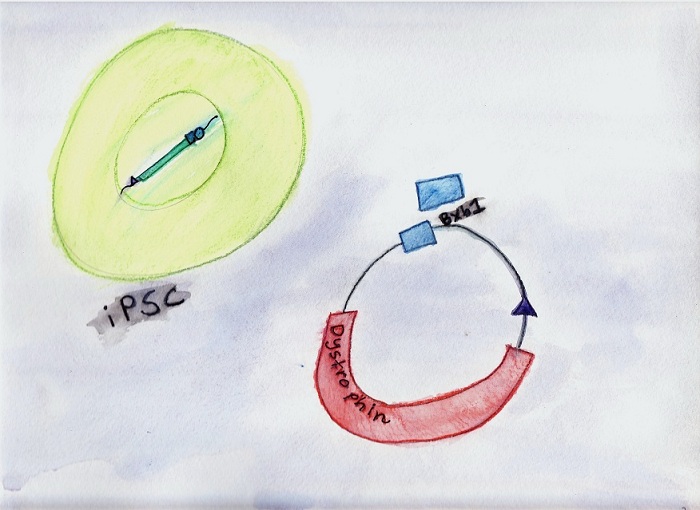
An area of intense interest in the lab is the use of genetically-engineered induced pluripotent stem cells (iPSC) to develop therapeutics. For example, iPSC derived from patients with genetic diseases like muscular dystrophy can be corrected, differentiated, then engrafted back to the patient.

We have used phiC31 integrase to add genes to reprogram adult cells into pluripotent stem cells:

  • Karow, M., Chavez, C.L., Farruggio, A.P., Geisinger, J.M., Keravala, A., Jung, W.E., Lan, F., Wu, J.C., Chen-Tsai, Y., and Calos, M.P. (2011) Site-specific recombinase strategy to create iPS cells efficiently with plasmid DNA. Stem Cells 29, 1696 - 1704. [Abstract]


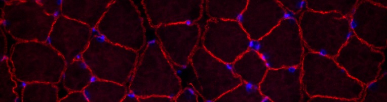
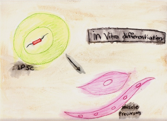
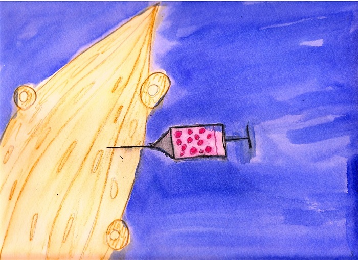
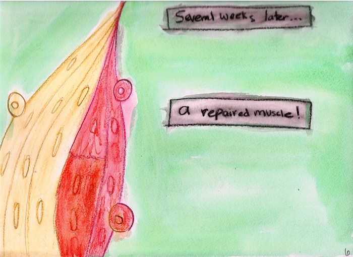
A current project involves reprogramming fibroblasts from mdx mice, a disease model for Duchenne muscular dystrophy, and adding the therapeutic dystrophin gene. Corrected cells are differentiated into muscle precursor cells in culture and are engrafted into mice to repair muscle damage.


This series of six images, created by Michele’s daughter Victoria, illustrates our first-generation system for reprogramming and gene correction:
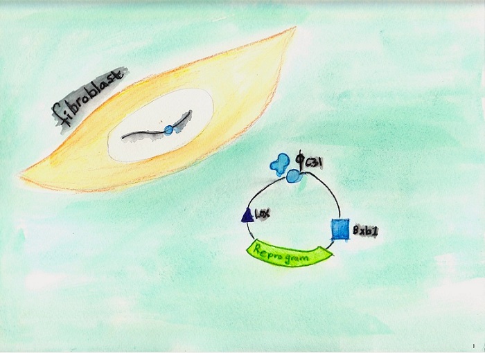

Victoria 1

1. Reprogramming.
In the first step, patient fibroblasts are reprogrammed by co-nucleofection with a plasmid carrying the four reprogramming genes (green region) and a plasmid encoding phiC31 integrase. The reprogramming plasmid also carries small recognition sites for recombinases: an attB site for phiC31 (blue circle, an attP site for Bxb1 integrase (blue square), and a loxP site for Cre resolvase (purple triangle). PhiC31 integrase (blue blob) pairs the attB site on the reprogramming plasmid with an endogenous pseudo attP site in the chromosomes (blue circle), to insert the reprogramming plasmid stably into the genome at a safe, intergenic location. Expression of the reprogramming cassette converts a subset of the fibroblasts into induced pluripotent stem cells (iPSC).


2. Correction.
The newly formed iPSC carry the reprogramming genes and an attP recognition site for Bxb1 integrase (blue square). To restore wild-type dystrophin to the iPSC, the cells are co-nucleofected with a plasmid carrying the dystrophin coding sequence, an attB recognition site for Bxb1 integrase (square), and a loxP site (triangle), together with a plasmid encoding Bxb1 integrase (square blob). The therapeutic dystrophin plasmid becomes precisely integrated at the Bxb1 attP site in the reprogramming plasmid resident in the chromosome.


3. Excision of unwanted sequences.
The top diagram shows a close-up of the sequences we have inserted into the chromosome. After reprogramming is complete, the reprogramming genes are no longer required and are detrimental to subsequent differentiation of the cells. Likewise, plasmid sequences that became integrated are no longer needed or desired. To remove the unwanted sequences, a plasmid encoding Cre is nucleofected into the iPSC. Cre causes precise recombination between two loxP recognition sequences that were engineered into the plasmid for this purpose. After excision, only the therapeutic dystrophin sequence, flanked by small recombinase recognition sites, remains in the iPSC.


4. In vitro differentiation.
The engineered iPSC are grown in culture under conditions that stimulate differentiation of a significant portion of the cells into muscle precursor cells. Such cells will bind to a labeled monoclonal antibody that recognizes muscle precursor cells, which include satellite cells, the normal stem cells resident in muscle tissue. Fluorescence-activated cell sorting (FACS) is used to isolate the muscle precursor cell population from other cells in the differentiating culture.


5. Intramuscular injection.

The sorted fraction of the differentiated iPSC containing muscle precursor cells is loaded into a syringe. The cells are injected into the tibialis anterior hind limb muscle of a mouse to assess engraftment of the engineered and differentiated iPSC.


6. Engraftment.
Injected muscles are analyzed three weeks or more after injection to evaluate whether engraftment has occurred. Engraftment can be detected in several ways. Immunocytochemistry performed on tissue sections can be used to detect the expression of dystrophin in the fibers. Detection of dystrophin staining in muscles that received engineered muscle cells compared to uninjected muscles suggests that engineered and corrected iPSC have engrafted in the muscle. The inserted dystrophin gene can also be detected in DNA isolated from engrafted muscle by using PCR, and dystrophin mRNA expression can be detected by RT-PCR.


Maintained by Tawny Neal
Last updated on Feb 14th 2013