About this Site
This site was created by Stacey Kallem and Kristyn Bigback for Humans and Viruses at Stanford University taught by Prof. Bob Siegel in Autumn of 2005.
Classification and Taxonomy:
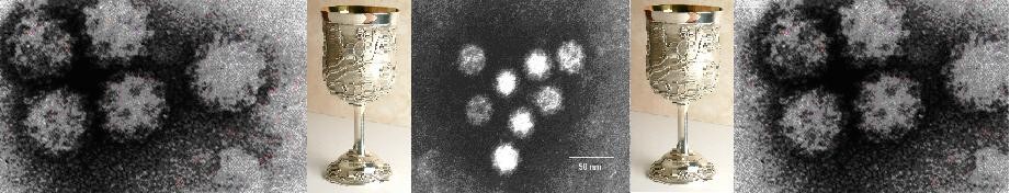
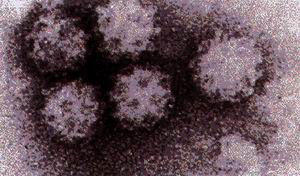
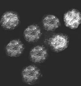
The name Caliciviridae comes from the Latin word for cup or chalice because there are 32 cup-like depressions in the surface of the virion when viewed in an electron microscope. This family has two genera that infect humans: the “Norwalk-like viruses” and the “Sapporo-like viruses.” The Caliciviridae family includes the genera Lagovirus and Vestivirus, which do not infect humans and thus will not be discussed in this webpage.
Virion:
Calicivirus virions are small (27 to 40 nm), icosahedral, and nonenveloped. They are resistant to detergents, drying, and acid (which allows it to survive in the acidic stomach environment to cause gastroenteritis).
Genome:
Calicivirus genomes are positive-sense, linear, nonsegmented, single-stranded RNA. They range from 7.3 to 8.3 kilobases in length, excluding the poly(A) tail. Nonstructural proteins are located at the 5’ end, and structural proteins are located at the 3’ end. All calicivirus genomes begin with a 5’-end terminal GU sequence (VPg), which is repeated internally in the genome and most likely corresponds to the beginning of a subgenomic-sized RNA transcript that is co-terminal with the 3’ end of the genome and that has been observed in infected cells as well as packaged into virions. The synthesis of at least one subgenomic mRNA in calicivirus-infected cells is a major difference between the replication strategy of caliciviruses and picornaviruses. Most calicivirus genomes contain three open reading frames (ORFs).
Transcription:
As with most positive-sense single-stranded RNA viruses, calicivirus genomes is first copied into a full-length negative-sense strand, which is then copied into full-length positive-sense
Pathogenesis:
Caliciviruses infect the intestinal brush border, preventing proper absorption of water and nutrients. This leads to diarrhea, vomiting, abdominal cramps, nausea, headache, malaise, and fever.
Transmission:
Fecal-oral route from contaminated water and food, especially shell-fish.
Prevention:
Prevention of calicivirus infection requires increasing sanitation and hygiene standards. This includes frequent and thorough hand washing, water sanitation techniques, as well as thorough and sanitary cooking of food.
No vaccine is available, and a major hindrance in vaccine development is the fact that the viruses themselves cannot be propagated as a source for live or inactivated vaccine. However, this problem may be circumvented by the availability of rVLPs, which are a replenishable and abundant source of capsid antigen
Treatment:
There are no antiviral drugs available for calicivirus infection. Calicivirus infection is usually self-limited and normally resolves on its own without complications. Oral fluid and electrolyte replacement therapy is usually sufficient to replace fluid loss. Parenteral administration of fluids may be necessary if severe vomiting or diarrhea occurs.
At-Risk Groups:
Children in day care centers, schools, resorts, hospitals, nursing homes, restaurants, cruise ships (due to infected food handlers).
Pictures cited from:
http://www.cornichon.org/archives/Shellfish%20platter%20at%20Cigale.jpg
http://www.caliciwatch.org/calici.jpg
http://www.ela-iet.com/images/REWbug4.jpg
http://www.ncbi.nlm.nih.gov/ICTVdb/WIntkey/Images/em_calic.gif
http://www.silverqueen.com/sept2003/Audubon%20Chalice.jpg
This site was created by Stacey Kallem and Kristyn Bigback for Humans and Viruses at Stanford University taught by Prof. Bob Siegel in Autumn of 2005.