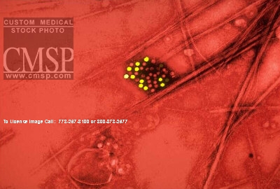
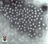
 Stool sample from an individual with gastroenteritis.
PHOTOS
Stool sample from an individual with gastroenteritis.
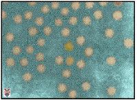
 An
astrovirus image from the ICTVdB.
An
astrovirus image from the ICTVdB.
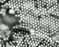 micrograph
prepared by Dr Hans Ackermann, Department of Microbiology, Laval University,
Quebec, Canada. Specimen stained with 2 % PTA . The bar represents 100 nm. Primary
magnification 29,700 x
micrograph
prepared by Dr Hans Ackermann, Department of Microbiology, Laval University,
Quebec, Canada. Specimen stained with 2 % PTA . The bar represents 100 nm. Primary
magnification 29,700 x
 A colorized electron micrograph from Prof. Stewart McNulty, Veterinary Sciences,
Queen's University, Belfast, UK.
A colorized electron micrograph from Prof. Stewart McNulty, Veterinary Sciences,
Queen's University, Belfast, UK.
 Method: Negative-stain Transmission Electron Microscopy. From the EPA's Microbiology
Website.
Method: Negative-stain Transmission Electron Microscopy. From the EPA's Microbiology
Website.
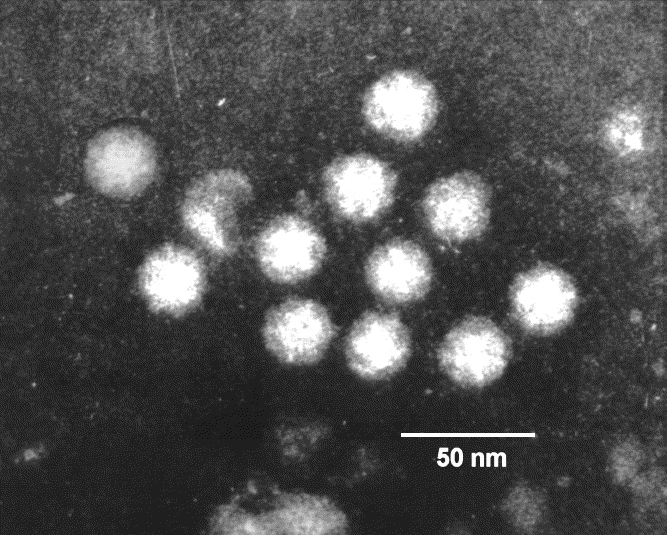
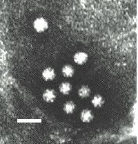 Note
the star-like images exhibited by individual virus particles. These are distinct
from the 'Star of David' image exhibited by typical calicivirus particles. Bar
= 50 nanometers.
Note
the star-like images exhibited by individual virus particles. These are distinct
from the 'Star of David' image exhibited by typical calicivirus particles. Bar
= 50 nanometers.