1994 Project Reports | Contents | Previous | Next | Home |
Hip / Acetabulum computer models to study prothesis design
JA Mandell, MS; ME Levenston, MS; Gary S Beaupré, PhD; SB Goodman, MD; David J Schurman, MD; Dennis R Carter, PhD
Overview - Joint degeneration due to arthritis or trauma adversely affects the quality of life of many older Americans. Over 200,000 patients are treated yearly with artificial joint replacements. While these operations have been very successful in restoring pain-free function, complications can occur, including prosthesis loosening, infection, inflammation, and bone or prosthesis fracture. The design of the components for joint replacement surgery can greatly influence the eventual success or failure of the procedure. At the Rehab R&D Center, we apply our engineering expertise to investigate how various aspects of component designs can be improved.
Many current joint replacement components are implanted without the use of bone cement. Porous surface treatments allow bone tissue to grow into the prostheses in order to provide stable long-term fixation. One area of concern for these joint replacements is relative motion (micromotion) between the prosthesis and the bone in the immediate post-operative period. If too much micromotion is produced when the patient uses the joint, the bone will not grow into the prosthesis. Instead of stable fixation, a fibrous (scar-like) tissue may develop which can lead to pain and eventual failure of the procedure. A second area of concern is long-term bone adaptation around the prostheses. When a natural joint is replaced with artificial components, the way the remaining bone supports the loads generated during daily activities is changed. Bone hypertrophies with increased loading and atrophies under decreased loading. Joint replacements generally result in reduced loading of the bone adjacent to the joint. This can lead to excessive loss of bony support for the prosthesis and subsequent prosthesis loosening (Figure 1).
 Figure 1. X-rays of a femoral prosthesis. Compare the bone immediately
after surgery (left) and after the prosthesis has induced bone loss (right).
Figure 1. X-rays of a femoral prosthesis. Compare the bone immediately
after surgery (left) and after the prosthesis has induced bone loss (right).
Among the techniques we use to study joint replacements is computer simulation of bones and artificial components. The most common joint replacement surgery is total hip replacement, which involves both sides of the ball and socket joint at the hip. The following sections describe two recent studies that used computer modeling to investigate micromotion and bone loss around artificial hip replacements. The first study examines the geometry of collars for components implanted in the femur (thigh bone), and the second examines the interface between acetabular (socket) components and the underlying bone in the pelvis.
Influence of Collar Geometry in Femoral Components
JA Mandell, MS; GS Beaupré, PhD; SB Goodman, MD; DJ Schurman, MD; DR Carter, PhD
In this study, we compared micromotion and load transfer among four prostheses: flat collared, 30 degree and 60 degree conical-collared, and tapered (Figure 2). According to our definition of collar angle, the flat collar and taper are equivalent to 0 degree and 80 degree conical collars, respectively. To isolate the effects of collar geometry, the implant models were straight-stemmed and cylindrical, and collar angle was the only design variable. Our goal was to quantify the relationship between collar angle, micromotion, and the details of load transfer in the early post-operative period.
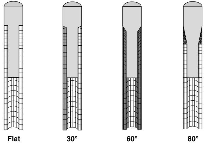 Figure 2. Computer models of femoral component collar geometries.
![]() The flat (0 degree)
collar used direct axial compression to transfer loads from the joint to the
adjacent bone. In contrast, the 30 degree, 60 degree, and 80 degree collars
transferred loads by "wedging" into the bone. Micromotion was lowest
in the flat collar model, due to the flat support provided by the bone, and
increased with collar angle, as increased wedging was accompanied by increased
sinking into the bone (Figure 3). Load transfer to the bone adjacent to the
joint increased with collar angle. The results suggest that femoral loading can
be maintained with conical-collared or tapered prostheses, but that increased
loading is accompanied by increased micromotion, which may prevent stable
long-term bony fixation. Current work includes efforts to predict long-term
bone adaptation around these prostheses.
The flat (0 degree)
collar used direct axial compression to transfer loads from the joint to the
adjacent bone. In contrast, the 30 degree, 60 degree, and 80 degree collars
transferred loads by "wedging" into the bone. Micromotion was lowest
in the flat collar model, due to the flat support provided by the bone, and
increased with collar angle, as increased wedging was accompanied by increased
sinking into the bone (Figure 3). Load transfer to the bone adjacent to the
joint increased with collar angle. The results suggest that femoral loading can
be maintained with conical-collared or tapered prostheses, but that increased
loading is accompanied by increased micromotion, which may prevent stable
long-term bony fixation. Current work includes efforts to predict long-term
bone adaptation around these prostheses.
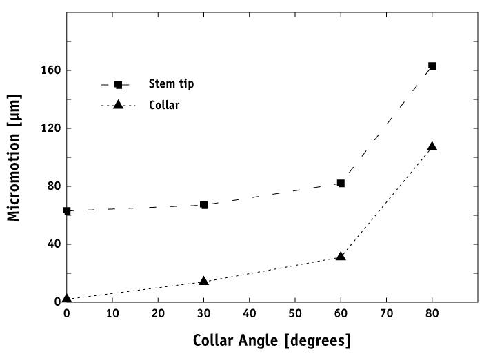
Figure 3. Micromotion of the prosthesis collar and the tip of the stem.
Bone Adaptation Around Acetabular Components
ME Levenston, MS; GS Beaupré, PhD; DJ Schurman, MD; DR Carter, PhD
An important consideration in the design of noncemented acetabular components is the achievement of component stability (little relative motion) in the immediate post-operative period. A low level of micromotion promotes growth of bone into the porous coating on the prosthesis, resulting in long-term fixation of the component. In this study, we compared the effect of this type of fixation on long-term adaptation. We also considered whether component features designed to produce post-operative stability have any direct influence on long-term bone changes.
We used a computer model representing a slice through the pelvis passing through the acetabulum and the points of contact with other bones (Figure 4). To predict long-term changes around the component, we applied a mathematical theory for bone adaptation developed by our research group. We considered two bounding cases for the condition of the implant/bone interface: a) a fully fixed prosthesis with bone growth into 100% of the porous coating and b) a fully loose prosthesis with no bone ingrowth (Figure 5). For a given interface type, we found that the different component designs produced virtually identical predicted adaptations. For all component types examined, the simulations consistently predicted extensive bone loss adjacent to the fully fixed components, indicating a high risk of eventual component instability or migration into the underlying bone. By contrast, the simulations with fully loose components predicted more moderate bone changes.
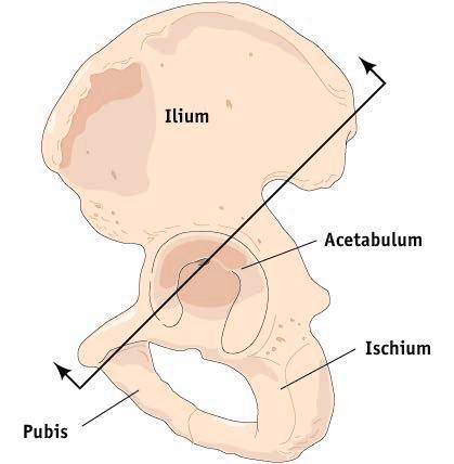
Figure 4. Diagram of the pelvis showing the orientation of the computer model. |

Figure 5. Results of the bone adaptation simulation for the a) fully-flexed and b) fully loose acetabular components. Areas of predicted bone loss (atrophy) and gain (hypertrophy) are shown in gray levels and patterns, respectively. |
Most current noncemented acetabular components have porous coatings over the entire prosthesis surface intended to produce maximal bony fixation. Our results indicate that this is not a beneficial long-term outcome. Based on this study, we suggest that porous coating be applied to selected areas of acetabular components to produce bony fixation only in desirable regions.
Republished from the 1994 Rehabilitation R&D Center Progress Report. For
current information about this project, contact
Gary Beaupré.