| Home | Projects | People | Publications | Places | | Arthritis | Osteoporosis | Stroke | Spinal Cord Injury |
Problem: Approach: Based on this result, a a 3-year clinical trial was begun in October, 1996, with Vincent R. Hentz, MD, Chief of Hand Surgery, as co-principal investigator; up to 10 patients per year requiring nerve repair in the hand will be enrolled starting in April, 1998. Related Work: Investigators:
Collaborators:
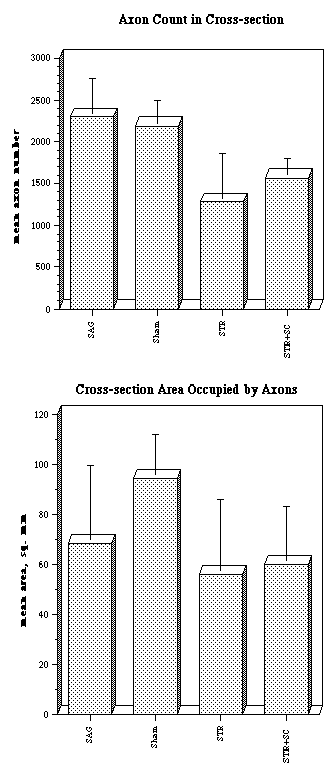
Findings from a 1994 experiment in which grafts containing oriented collagen core matrix with ("STR+SC") or without ("STR") rat Schwann cells were implanted into rat peroneal nerves; controls were sutured autografts ("SAG") and sham operations. Exposure of the nerve alone caused inflammatory response and some functional loss. Autografts and Schwann-cell seeded grafts recovered >90% of function after 3 months. Without Schwann cells, recovery averaged 70% in the same period. The bar graphs show group means of cross-sectional histology. There were higher numbers of myelinated axons distal to the implant in SAG and sham groups, including multiple branches as occur during the healing process. Area occupied by axons was also lower in both STR groups, a somewhat misleading measure, since most of the collagen core is still intact and displaces axons. The conclusion is that the high surface area of the core and the Schwann cell population promote rapid elongation and maturation (i.e.: elimination of excess branches) of axons through the implant to their target muscle or sensory connections (illustrated by the microphotograph of a core element with immunostained axons from an in vitro experiment). Last updated March 26, 1999 |