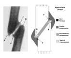
Investigators: Elizabeth G. Loboa, MS Project Category: Arthritis - 2000 Summary: Mechanical stresses play an important role in regulating tissue differentiation in a variety of contexts during skeletal development and regeneration. It has been shown that some intermittent loading at a fracture site can accelerate secondary fracture healing. However, it has not been shown how the stress and strain histories resulting from mechanical loading of a fracture might, in some cases, inhibit normal fracture healing and induce pseudarthrosis (false joint) formation. In this study, finite element analysis is used to calculate hydrostatic stress and tensile strain patterns in regenerating tissue around a transverse osteotomy and an oblique fracture. Using a mechanobiologic view on tissue differentiation, we compare calculated stress and strain patterns within the fracture calluses of both geometries to the histomorphologies of a healing transverse fracture and a typical oblique pseudarthrosis. Purpose: Mechanobiology is the study of how mechanical or physical stimuli regulate biological processes. It is clear that mechanobiology plays a critical role in fracture healing. The purpose of this study is to examine: 1) the role of tensile strains in the differentiation of pluripotential tissue at a transverse osteotomy and an oblique fracture site; and, 2) the role of hydrostatic pressure in the differentiation of pluripotential tissue at a transverse osteotomy and an oblique fracture site. Methodology: This study uses theoretical and computational models to investigate the role of mechanical stimuli in the differentiation of pluripotential mesenchymal tissue. Our theoretical model is based on the assumption that hydrostatic pressure directs the pluripotential mesenchymal tissue of a fracture callus down a chondrogenic pathway, significant tensile strain leads to fibrogenesis, a combination of hydrostatic pressure and significant tensile strain leads to fibrocartilage development, and, given adequate vascularity, low levels of hydrostatic stress and tensile strain allow direct intramembranous bone formation. It also incorporates the idea that excessive hydrostatic pressure can lead to pressure necrosis and excessive tensile strain can cause tensile failure of the mesenchymal tissue. Our computational models consist of two-dimensional finite element models of both a transverse osteotomy and an oblique fracture. These models are used to calculate hydrostatic stress and tensile strain patterns within the fracture calluses. These stress and strain patterns are then analyzed in the context of the mechanobiologic tissue differentiation concept presented above. Progress / Results: Finite element models of both transverse osteotomy and oblique fracture sites have been created and analyzed. Tissue differentiation predictions appear to be consistent with the characteristic histomorphologies of both healing transverse osteotomies and oblique pseudarthroses. The oblique pseudarthrosis model is being further developed into a three-dimensional model to better represent later stages of pseudarthrosis development. 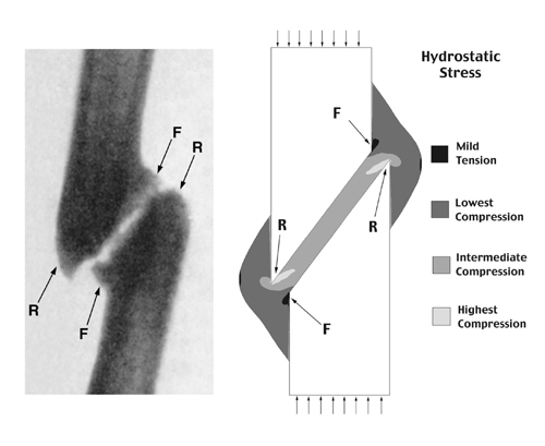 Caption: Oblique pseudarthrosis formation. Roentgenogram of an oblique pseudarthrosis. (Adapted from Pauwels F:. Biomechanics of the Locomotor Apparatus. Springer-Verlag, Berlin. p. 124, 1980.) Hydrostatic stress plot from a finite element analysis of compressive forces acting on an oblique fracture site. Fibrocartilage and cartilage formation occur in the interfragmentary gap, a region of hydrostatic compression. Bone formation occurs periosteally in areas of mild hydrostatic tension (marked "F") and bone resorption occurs in areas of high hydrostatic compression (marked "R"). Funding Source: VA RR&D Merit Review |