| Home | Projects | People | Publications | Places |
| Previous Story | Press | Next Story |

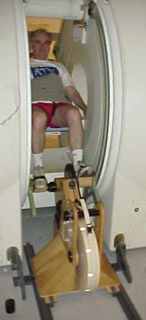
Custom MRI cycle tracks blood flow during exercise
From: Stanford Report - January 22, 2003 - page 7
By: Sara Selis
For most people, an MRI scan requires lying completely motionless while they are transported inside the narrow bore of the giant MRI magnet. At the medical center, however, a years-long collaboration among faculty in radiology, vascular surgery and mechanical engineering has made possible a completely different MRI experience in which the patient sits upright and pedals a custom-built exercise cycle inside the machine. The cycle, believed to be the first of its kind, enables School of Medicine researchers to precisely and non-invasively measure patients' blood flow while they exercise. That information yields a better understanding of vascular disease, which could ultimately lead to improved diagnosis and treatment. "If you want to understand patients' blood flow, you need to see them in various physiological states," explained Charles Taylor, PhD, assistant professor of surgery and mechanical engineering, who spearheaded the project along with radiology professor Robert Herfkens, MD. "Until now, we could accurately measure blood flow only when the patient was at rest, and with vascular disease, that's not enough." With some vascular disorders, symptoms become apparent only during physical activity. MRI technology has long been used to measure blood flow at rest, but obtaining such data on an exercising patient wasn't feasible for various reasons: The standard-model MRI's enclosed design made exercise there virtually impossible. No metal is permitted inside the magnet. And to ensure accurate readings, patients must remain still. Researchers at other institutions tried various methods to measure blood flow during exercise, but these carried draw-backs. One method involved inserting a catheter inside the femoral artery, but this approach was invasive and could temporarily alter the artery's function. Another strategy was to do an MRI scan on the patient immediately following exercise, but this yielded inaccurate results. When Stanford acquired a high-powered, vertically open MRI in 1997, Taylor and Hertkens saw an opportunity to custom-design an exercise cycle for it. They assigned the project to a Stanford engineering class, which built a rudimentary prototype mostly of wood. Following a second iteration, Chris Cheng, then a doctoral student in mechanical engineering, took on the project in winter 2000 as part of his thesis. Cheng supervised a group of engineers at the Palo Alto Veterans Affairs Rehabilitation Research and Development Center to design and construct a more durable, streamlined model. The final product, weighing about 80 pounds, consists of beech wood and high-density plastic.
Stanford researchers are using the cycle in basic research on healthy adults and patients with intermittent claudication, a condition in which blood flow to the legs is obstructed, causing stiffness and pain. The data from the healthy patients is helping researchers understand normal blood flow under various conditions. Data from the second group is shedding light on a common vascular disorder, which could lead to better diagnosis and treatments. "This bike is a tool that lets us squeeze more information from this already-useful [MRI] machine," said Ronald Dalman, MD, associate professor of vascular surgery at the Veterans Affairs Palo Alto Health Care System, who is using the cycle to monitor some of his patients. "In just a few minutes, you can get an incredible amount of useful data, which opens new possibilities for understanding vascular disease." In collaboration with Jeffrey Feinstein, MD, assistant professor of pediatrics, Stanford researchers will soon use the cycle to monitor blood flow in children treated for congenital cardiovascular disease. Herfkens said multidisciplinary collaboration has been key to the project's success. "Making it work requires fantastic imaging equipment, engineers to develop the right hardware and software, technicians to run the equipment, and, clinicians who want to use the data." |