1996 Project Reports | Home |
Contents | Previous |
Next |
Effect of hemiarthroplasty on acetabular cartilage
Marjolein C. H. van der Meulen, PhD; William A. Allen, BS; Virginia L.
Giddings, ME; Kyriacos A. Athanasiou, PhD; Robert D. Poser, DVM; Stuart B.
Goodman, MD, PhD; R. Lane Smith, PhD; Gary S. Beaupré, PhD
One third of all hip replacements performed in the U.S. annually are
hemiarthroplasties. A hemiarthroplasty, as opposed to a total hip replacement,
involves the replacement of the femoral side of the hip joint by a rigid metal
implant which rests in the patient?s acetabulum against the natural cartilage
(Fig. 1). Compared to a total hip replacement, hemiarthroplasty procedures
involve shorter surgical times and lower medical and prosthesis costs. Cost
considerations make it likely that the number of hemiarthroplasties performed
will increase in the future. Hemiarthroplasty success, however, is often
limited by pain from extensive cartilage erosion and loss of joint space. The
severe cartilage degradation resulting from articulation with a metal implant
is of great concern if hemiarthroplasty is to be a more successful surgical
procedure.
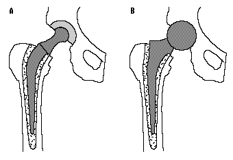 Figure
1. Schematics of (a) total hip arthroplasty with prosthetic replacements of
both sides of the hip joint; (b) hemiarthroplasty with femoral prosthesis only.
The purpose of the present research is to fully characterize the changes
that occur in the acetabulum following hemiarthroplasty, and to also examine
surgical variables which may contribute to this degradation. We hypothesize
that the altered loading environment created by the introduction of the rigid
spherical prosthesis head will result in cartilage changes in the acetabulum.
|
We examined the morphological, biochemical, material and histologic changes
of acetabular articular cartilage after one year of hemiarthroplasty in
nineteen sheep. Visual examination of the left acetabulum determined the extent
of cartilage erosion (% loss of cartilage area) and the condition of the
remaining cartilage (% fibrillation). Cartilage degradation was assessed
biochemically by examining the primary constituents of cartilage: proteoglycans
and collagen. Material properties and thickness were determined from creep
indentation experiments. Histologic appearance was graded from thin sections.
Differences between the acetabulum pairs were analyzed by paired t-tests.
From visual examination, twelve sheep demonstrated moderate loss of
cartilage (10-50%), and seven sheep had widespread cartilage loss (50-90%). In
fifteen sheep, the remaining cartilage exhibited widespread or complete
fibrillation. Both the biochemistry and the material property data indicate
severe degradation of the remaining cartilage. We are currently in the process
of confirming these results with the histology. Upon completion of the
cartilage characterization, we will examine the correlation of the degradative
changes with surgical parameters, including head size, leg length, and neck
angle.
Republished from the 1996 Rehabilitation R&D Center Progress Report. For
current information about this project, contact:
Marjolein C. H. van der Meulen.
