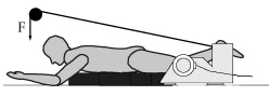
Figure 1. Typical set-up for patellar study. Subject raises a load in extension.
1996 Project Reports | Home | Contents | Previous | Next |
Frances Sheehan, MS; Felix E. Zajac, PhD; John Drace, MD
Objective - An understanding of the way in which the neurological, muscular and skeletal system work together to complete a specified task has long been a major goal of experimental and modeling studies. Unfortunately, most kinematic and kinetic properties of the musculoskeletal system cannot be measured directly, without the use of invasive techniques. Human studies are typically limited to inferring muscle and bone kinematics and kinetics from externally measured quantities such as EMG (neural excitation), joint torques, and skin marker positions . This study has been directed at adapting the presently available Cine Phase Contrast Magnetic Resonance (PCMR) Imaging techniques to the study of the human musculoskeletal system during a dynamic task. Cine PCMR was originally developed to image heart motion and blood flow. For each time frame, PCMR provides an anatomic (magnitude) image and x, y and z velocity images, or velocity maps (VMs). The magnitude image shows the familiar gradient echo contrast between tissues. The VMs depict the 3-D velocity vectors of all points within the imaging plane.
A major goal of this study is to use the 3-D VMs to track bone and muscle movement during a particular motion cycle in the hope of developing clinically relevant diagnostic MRI tools. Recent work has focused on characterizing patellar tracking within the femoral groove in a set of healthy volunteers and a set of patients diagnosed with patellar tracking problems. Besides the bony motion, the muscle kinematics and kinetics of the knee extensors, which control patellar motion, has been studied. Other studies of interest include characterizing muscle motion before and after muscle-tendon transfers in the forearm, and characterizing muscle adaptation after disuse resulting from immobilization or denervation. All these studies have the common theme of musculoskeletal changes in response to an injury. A better understanding of such changes may facilitate rehabilitation.
| Figure 1. Typical set-up for patellar study. Subject raises a load in extension. |
Approach - In order to test the accuracy of Cine PCMR as it relates to the study of dynamic musculoskeletal motion, a motion phantom, consisting of a series of paired plastic gears connected by a plastic rods, was developed to move rigid samples in complex 2-D and 3-D trajectories. The samples consisted of CuSO4 gelatin solution or trabecular bone with fiducial markers. The rods of the phantom could be adjusted to vary in distance and orientation to simulate complex deformation of muscle samples. Trajectories of deformable or rigid regions of interest (ROIs) in the phantom experiments were tracked using the VMs and were compared to the ROIs' paths, calculated from the paths of the fiducials, as observed on the magnitude images.
To date, forearm muscle motion has been studied in 8 controls and 5 patients. The patients were either undergoing tendon transplant or had experienced denervation of some of the muscles of the forearm. Knee (patella and vasti muscles) studies have been performed in 6 patient and 12 control knees during active leg extensions/flexions. The patients had been diagnosed with patellar tracking problems, which caused pain and limited the use of the joint. Soleus/gastroc measurements have been undertaken in 6 controls, in the hope that this data can be used as a comparison to data from subjects who have experienced long term dissuse of the muscle.
Conclusions - Measurements of tendon stretch, muscle deformation, translation, rotation, stress and shear using PCMR are feasible. The phantom study showed that the errors in tracking both bone and muscle motion were minimal (approximately 0.5 - 1.4 mm).
In early clinical feasibility tests, the ability of Cine PCMR to distinguish normal forearm muscle kinematics/kinetics from those of denervated muscle was dramatic. Preliminary data on vasti motion discrepancies may play a role in assessment of therapy of patients with patellar tracking abnormalities. The preliminary work on patellar tracking shows similar values for specific clinical angles (such as patellar tilt angle) as reported in the literature.
 Figure 2. Displacement (mm) of an ROI on the belly of the Vasti Lateralis muscle during active knee extensions / flexions under different loads. The weight was raised during extension. Velocity maps in the calf and thigh show substantial difference under different conditions of force applied. Technical developments may allow improved temporal resolution and 3-D volume data capability but new analysis techniques need to be conceived to comprehensively understand velocity distributions in muscle tissue. |
Republished from the 1996 Rehabilitation R&D Center Progress Report. For current information about this project, contact: Frances Sheehan.
![]()