1996 Project Reports | Home | Contents | Previous | Next |
Marjolein C. H. van der Meulen, PhD; Marienne Moro, MS; Marvin W. Ashford, MS; Joanne DeAussen, BS; Amanda Carter, BS; B. Jenny Kiratli, PhD; Robert Marcus, MD; Laura K. Bachrach, MD; Dennis R. Carter, PhD
Dual energy x-ray absorptiometry (DXA) has gained widespread acceptance in the clinic for measuring bone mass. This technique can measure bone mineral content, bone width and bone area in the scan plane, but cannot measure the true cross-sectional geometry and volumetric density of a bone (Fig. 1a, b). At a given cross-sectional location, the mineral content through the depth of the scan is measured, but the actual geometric arrangement of this material in the cross section of the bone cannot be determined. Knowing not only the total bone mass but also its distribution is important to understanding bone structural behavior.
Figure 1. Three representations of a femur midshaft cross section: shown schematically as a) actual shape, b) calculated mineral distribution curve, and c) idealized circular model
Most bones of the skeleton serve an essential structural role in the body, and to evaluate their ability to perform this role requires knowledge of the geometry as well as the material properties. Vertebral osteoporotic fractures, for example, are the result of structural changes that weaken the spine and failure occurs when the structure can no longer bear the loads during daily activities. Therefore, a structural assessment based on DXA measurements is desirable. Previous researchers have shown that if the cross-sectional shape is assumed to be a hollow circle of a constant material density, the DXA output curve can be mapped onto this circular section (Fig. 1c). For the dense cortical bone of the mid-shaft, this is an appropriate assumption, and allows us to construct a structural model directly from the DXA data.
Our research has used this structural approach to examine femoral bone mass obtained from DXA measurements for several different populations. Our goal is to understand the factors that influence the structural properties of the skeleton. The populations being studied are of particular interest in the study of osteoporosis: adolescents and individuals with paralysis following spinal cord injury. Adolescence is a time of rapid bone growth and much of our peak adult bone mass is acquired at this stage of life. Following spinal cord injury, the loading to the lower extremity is dramatically altered.
The amount of bone gained during adolescence along with the subsequent rate of bone loss have a profound impact on an individual's total bone mass in adulthood and old age. At least 50% of our peak adult bone mass is gained during adolescence when dramatic changes in body size and structure occur. Many different factors influence bone growth including age, gender, ethnicity, body mass, pubertal stage, and hormones. However, the exact role and relative contributions of these different influences are unknown.
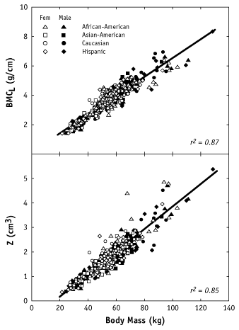
In a population of male and female adolescents and young adults (aged 9 - 26 years) from different ethnic backgrounds, our goal was to examine several likely determinants of bone cross-sectional geometry and structure including age, pubertal stage, body mass, and height. Our sample consisted of 97 African Americans (56 females/41 males), 97 Asian Americans (48/49), 101 Caucasians (53/48), and 80 Hispanics (45/35). Height and body mass were measured, and pubertal stage was determined by self-assessment. Whole body DXA scans were used to determine the diaphyseal length and mid-diaphyseal diameter of the left femur, as well as the bone mineral content of a region at the mid-shaft. Based on the hollow circular model described above (Fig. 1), cross-sectional geometric properties (area, moment of inertia) were estimated and used to calculate two structural strength indicators: the section modulus and the whole bone strength index. For the measured bone parameters, we first examined simple linear regressions with age, pubertal stage, body mass, height, and lean and fat mass. Next, gender and ethnicity were added to the linear regressions to see if additional predictive power was provided. Finally, a saturated model including all possible determinants was tested.
The relationships between the bone measurements and age, pubertal group, height or body mass showed the strongest correlations with body mass (Fig. 2). Accounting for gender and ethnicity provided little additional predictive value over the simple regressions with body mass alone. Furthermore, taking all developmental parameters into account in a single regression did not significantly improve the predictive power obtained using only body mass.
|
Figure 2. Regression analyses of bone mineral density (BMD) and section modulus (Z) on body mass for children and adolescents of four ethnic groups. These results indicate that increases in mid-femoral bone mass and cross-sectional properties during adolescence are primarily related to changes in body mass. We believe that body mass is an indicator of in vivo loading and that the strong relationship between body mass and mid-femoral bone structure is due to increased mechanical loading. At present, we are applying a similar approach to the femoral neck. Due to the prevalence of osteoporotic fractures, the femoral neck is a site of considerable clinical interest. The geometry is very different from the mid-femur, and the structure consists not only of dense cortical bone, but also contains a trabecular core. We would like to understand developmental factors that determine the changes in structure. |
Republished from the 1996 Rehabilitation R&D Center Progress Report. For current information about this project, contact: Marjolein C. H. van der Meulen.
![]()