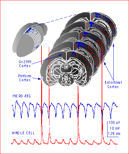
 Theta activity can be recorded
from specific regions of cortex (blue dots) in
rat brain slices. Comparison of micro-EEG
signals and intracellular recordings (whole cell) reveal that the low frequency
theta waves (~ 8 Hz) were generated by synchronous synaptic potentials and
discharge activity of cortical neurons. The discharge of each cortical neuron
appears to contribute ~ 1.0 µV to the micro-EEG signal, so theta activity
requires synchronous activity in ~ 100 neurons in each recording location.
Theta activity is known to be important for spacial mapping and may
provide a 'binding' mechanism that contributes to the formation of memory
in general. When selective populations of neurons are synchronously active
they can interact in a Hebbian manner to change the strength of synaptic
inputs that are timed at the theta frequency. Theta activity is also known to be particularly sensitive to anesthetic
agents at concentrations which block memory formation.
Theta activity can be recorded
from specific regions of cortex (blue dots) in
rat brain slices. Comparison of micro-EEG
signals and intracellular recordings (whole cell) reveal that the low frequency
theta waves (~ 8 Hz) were generated by synchronous synaptic potentials and
discharge activity of cortical neurons. The discharge of each cortical neuron
appears to contribute ~ 1.0 µV to the micro-EEG signal, so theta activity
requires synchronous activity in ~ 100 neurons in each recording location.
Theta activity is known to be important for spacial mapping and may
provide a 'binding' mechanism that contributes to the formation of memory
in general. When selective populations of neurons are synchronously active
they can interact in a Hebbian manner to change the strength of synaptic
inputs that are timed at the theta frequency. Theta activity is also known to be particularly sensitive to anesthetic
agents at concentrations which block memory formation.
Theta activity requires complex circuit interactions
between cortical neurons and appears to be more
sensitive to anesthetics than single
monosynaptic responses. We expect to find that anesthetics act at multiple
pre- and postsynaptic sites to disrupt
the synchronous activity of circuits of neurons.
Other brain slice EEG labs: Avoli at McGill, Bland at U
Calgary, Buzsaki
at Rutgers, Hasselmo at
Boston U, Randall
at GlaxoSmithKline, Antkowiak
at U Tuebingen, McBain at NIH,
Reyes at New York
U, Whittington
at U Newcastle, Konopacki
at U Lodz, Sejnowski at the
Salk Inst., MacVicar
at UBC, Gahwiler
at U Zurich, Kauer
at Brown U, Alger
at U Maryland, Lynch
at UC Irvine -- let me know if I've forgotten anybody please.