Stanford


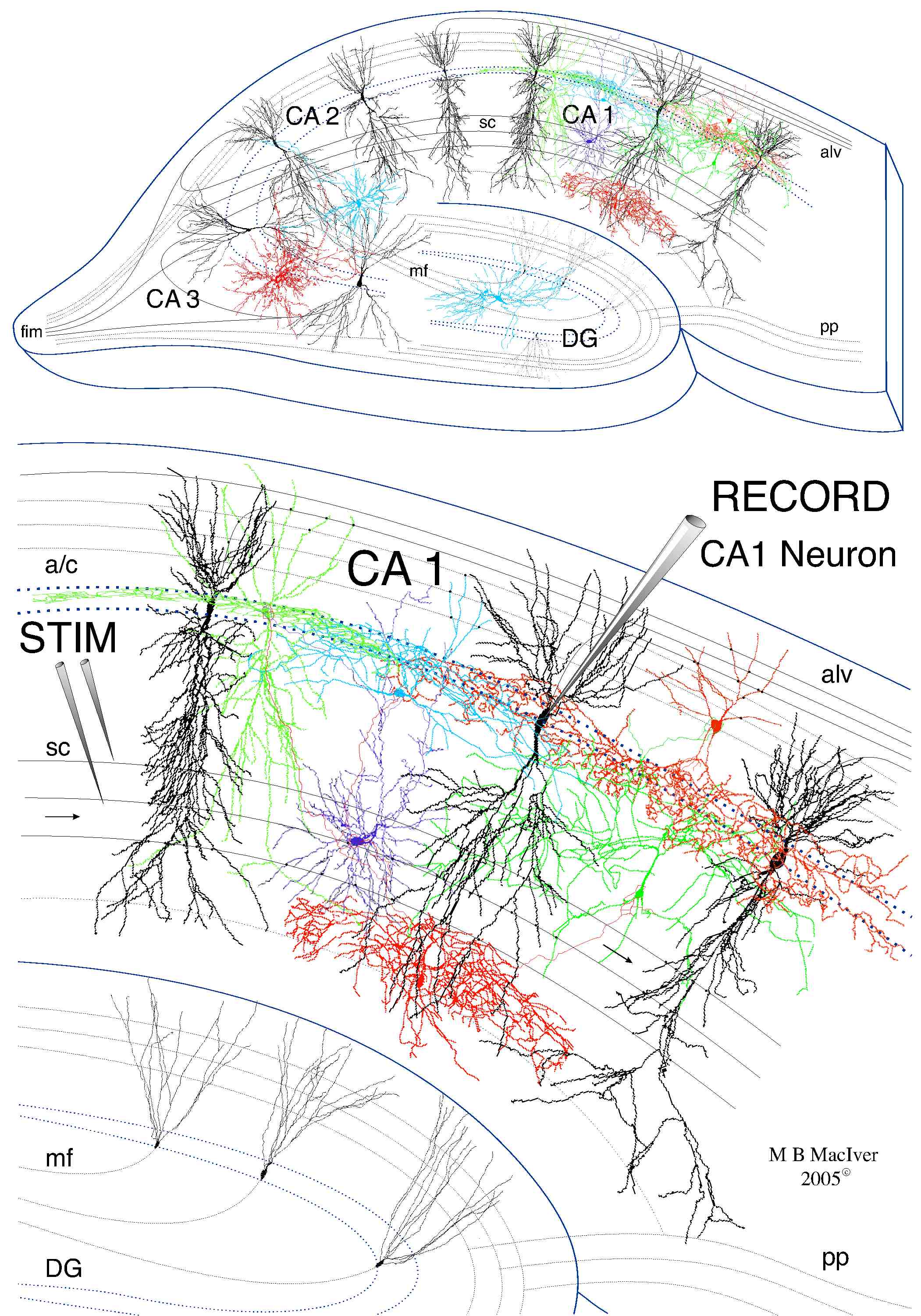
Hippocampal CA 1 pyramidal neurons in brain slices provide an ideal model system for studying drug effects on synaptic transmission. Intact synaptic pathways and interneuron circuits can be preserved in thin slices (0.4 to 0.5 mm) of brain tissue which remain viable for up to 24 hours. Brain slice circuitry for the CA 1 area is shown below:


Hippocampal Slice


Projection neurons and main synaptic pathways are shown in black, inhibitory interneuron colors represent cells that function in concert (red) vs  cells that belong to different inhibitory classes.  By activating the appropriate interneuron class, an input can selectively control large groups of projection cells.  Thus interneurons can gate or index groups of output cells, like CA1 pyramidal neurons, so that subpopulations can be synchronously active, e.g. during theta, and/or gamma frequency EEG oscillations.  Abbreviations: CA1 - cornu ammonis region # 1 of the hippocampal formation; STIM - stimulating electrode used to activate excitatory and inhibitory inputs to CA1 neurons; RECORD - recording electrode to measure pyramidal neuron response to stimulation; a/c - association/commissural pathway from septum and contralateral hippocampus; alv - alveus; sc - Schaffer collateral pathway from CA3 neurons; mf - mossy fiber pathway from dentate gyrus (DG) granule neurons; pp - perforant path axons from entorhinal cortex; fim - fimbria pathway to and from midbrain and other regions.


Neuropharmacology Home Page