Audition - Spiking precisely
Barn owls catch mice scurrying in the dark by telling which ear sound arrives at first—to within a microsecond! The brain's sound localization trick relies on the coincidence of precisely timed excitatory and inhibitory inputs, with excitation leading in one ear and inhibition in the other.
By modeling potassium channels found in auditory neurons, we have developed silicon neurons that lock on to acoustic features.
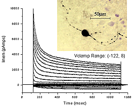
Low-threshold potassium channels open when the membrane voltage is below the threshold for spiking; they prevent the voltage from increasing further by shunting synaptic inputs. But these channels open with a delay of a few milliseconds. Thus, the large number of synaptic inputs received at onsets and peaks can raise the membrane voltage fast enough to outrace the potassium current and produce a spike with negligible delay.
From its detection in the ear to its perception in the cortex, sound is encoded with extreme precision.
Sound is separated into its constituent tones by the cochlea, a spiral structure in the inner ear. The cochlea's basilar membrane vibrates at its base when stimulated by high frequencies and vibrates at its apex when stimulated by low frequencies; the location progresses smoothly from base to apex as frequency drops. Spike discharges in the auditory nerve signal these vibrations to the brain, reporting them moment by moment, up to a few thousand times per second.