.....
.....
.....
.....
.....
.....
.....
.....
.....
.....
.....
.....
| ..... ..... ..... ..... ..... ..... ..... ..... ..... ..... ..... ..... ..... |
|
(The "![]() "
symbols are links. For problems or questions contact Wine.)
"
symbols are links. For problems or questions contact Wine.)
.
This is the Edwin
Smith surgical papyrus. It is a copy, made
about 3000 years ago, of a much older original that was written in the
Old Kingdom of Egypt, at the very dawn of history, 5000 years ago. I show
it to you for two reasons. Its existence illustrates a profound innovation
in the evolution of animals. The human brain, uniquely among all animals,
can create and interpret lasting symbols, and in this way can communicate
across vast reaches of space and time.
This ability is unprecedented in evolution. Prior to humans, animals only passed on their genes. Humans can pass on both their genes and their ideas. However, because passing on ideas is a late evolutionary innovation, the process is not yet perfected. It is not as efficient as passing on genes, and it is certainly not as enjoyable.
What is this ancient Egyptian saying to us? The papyrus was written by a surgeon, who was describing a gaping head wound in a soldier which gave the surgeon an exceptional opportunity to view a living brain. This papyrus is an icon to neuroscientists because it contains the oldest known reference in any written language to the brain. These words here mean brain, they are literally translated as the marrow of the skull, and in spite of its historical significance, the papyrus contains no special secrets For all of their wisdom, the Egyptians had no conception of the brain's importance, and embalmers routinely drained it out through the nostrils and discarded it, while keeping for mummification such internal organs as the liver and the spleen.
My job today, is to give you a brief introduction to a biological and genetic approaches to psychology.
1. EVOLUTIONARY CONCEPTS ARE ESSENTIAL.2. LANGUAGE ® CULTURE ® NEW BALLGAME.
3. BRAINS ARE MACHINES THAT THINK.
4. BRAINS ARE MADE OF NEURONS.
5. NEURONS ARE EXTREMELY COMPLEX CELLS.
6. NEURONS ARE ORGANIZED INTO NETWORKS.
7. BRAIN STRUCTURE IS LARGELY INHERITED.
8. STRUCTURE ® FUNCTION.
This is the chunk of tissue we are concerned with. This is a photograph of a preserved human brain. It weighs about 3 pounds, and is 78% water, 10% fat, 8% protein, and 4% other. There isn't much magic in those figures. But to express the brain in such reductionistic terms misses everything important. For this prosaic looking organ is an electrochemical machine, and it is the most highly structured material that we know about in the entire universe. The Pentium 4 processor from Intel has ~42 million transistors on one chip. A single neuron is vastly more complicated than a transistor, but even if we consider them equivalent, your brain has ~ 300 times more neurons than the Pentium processor has transistors. However, transmission speeds in the chip are roughly a million times faster than your neurons, and the Pentium 4 can carry out 1.7 billion instructions each second. (J. Markoff, N. Y. Times, 6-10-2001). As computers approach the size of brains, their vastly faster procesing speeds will allow them to perform better than human brains in a wider array of tasks. How far this process will go is a matter of great interest.
The goal of neuroscience is to understand this machine. But is it important for psychologists to think about the brain, and to incorporate biological concepts into our theories. Some psychologists have argued that it is not important to understand the material of the brain, anymore than it is important to understand the material of a book, because psychology is about symbolic systems, and equivalent symbolic systems can be represented in different material systems. What do you think about that?
One way to begin to analyze the brain is to cut it open and look at its parts. This shows a section right down the sagittal plane of the brain that divides it neatly into two halves. Here, we can see most of the major structures of the brain: the cerebral cortex has the greatest mass in a human brain. This prominent structure is called the cerebellum or little brain. This long structure is actually a huge, flattened internal nerve tract, cut in cross section. It is called the corpus callosum, and it serves to convey information back and forth between the left and right halves of the brain. All of the structures in this deep, middle portion of the brain are well developed in vertebrates down through reptiles. I won't list the numerous structures here, except for three: this apparently insignificant region is the thalamus, which is a major processor for information entering and leaving the cortex and cerebellum. Just below it is the even smaller hypothalamus, which, via neural connections with the brain and hormonal connections with the pituitary gland, can mediate interactions between our mental processes and such fundamental biological processes as sexual development and growth. Finally, we come to the spinal cord, which is both a massive tract of nerves and a series of local brains: you can think of the spinal cord as a string of miniature brains, each one mainly concerned with a horizontal segment of the body, with which it communicates via nerves.
One remarkable thing that we have learned about the brain is that all of the information that enters and leaves the brain travels as digital electrical signals over highly organized cables called nerves. That is one of those remarkable facts that everyone knows but no one fully appreciates or comprehends. Think of it: all of your experience of the world, all of the richness and nuance, all of the flavors and pains, originates as a series of simple signals that have no more individuality than the pulses of electricity in a telegraph line. Although each line is simple, a great many of these lines travel into and out of your brain. Every cubic millimeter of your body is invaded by many nerves, which carry information from hundreds of millions of receptors and convey, over about 5 million output lines, messages to every muscle and gland. If all of the tissue of your body except your nerves were to suddenly disappear, the remaining nerves would form a gossamer image of your body.
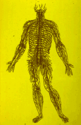
| This is a famous woodcut, remarkable both as art and as science. It was published in 1530 by the noted anatomist Vesalius, but the actual drafting may have been done by Titian, who was reputed to be one of Vesalius assistants. The combination of grace and power is certainly unusual in an anatomical drawing. It depicts the major nerves of the body, which are considerably exaggerated in thickness to aid the eye. How is information conveyed over these nerves? | |
This shows one idea. In 1662, Descartes proposed an essentially mechanical description of reflexive behavior. The only well-developed physical analogies that Descartes knew about were heat and hydraulics, and so he used them. Descartes proposed that when we jerk our hand or foot away from a fire, it is because the stimulus sets in motion animal spirits, contained in tubular nerves. The disturbance is conveyed to the ventricles of the brain where it prompts the pineal gland to release animal spirits into the nerve that runs to the associated muscle. The muscle inflates, and this inflation is mechanically coupled to the limb in such a way as to withdraw it.
Descartes concept has many important features that are not obvious in this simple drawing. Most important is his insistence that physical events intervene at every step between stimulus and response. More technically, Descartes recognized the need to convey peripheral signals to a central point, so that the actions of various limbs could be coordinated. Even some of the fine and apparently incidental features of the model turn out to be correct: neural messages really are conducted along hollow tubes, but the physical nature of the message is much more subtle and infinitely more complex than even a genius such as Descartes could have imagined, given the limitations of science in the 17th century. How does it really work?
This shows a modern view of a simple reflex arc, consisting of a muscle, the spinal cord, and two connecting nerve cells. A real nerve is a bundle of nerve cell axons. Each axon is indeed a hollow tube, but they are exceedingly fine and numerous: they are thinner than hairs, and a single thin nerve contains thousands or tens of thousands of axons. The message that travels along a single axon is called a nerve impulse.
This reflex arc mediates the stretch reflex. When this muscle is stretched, the mechanical deformation is converted by a stretch receptor into nerve impulses. This process is called transduction. The nerve impulses travel along the axon of the sensory nerve cells (only one is shown here), and, when they reach the terminal of the sensory nerve cell in the spinal cord, the nerve impulses causes the terminals to secrete chemicals which in turn stimulates other neurons in the spinal cord, including motor neurons. This process is called synaptic transmission. If synaptic transmission is strong enough, the motor neuron generates nerve impulses, which are conducted along the nerve to the muscle by a mechanism identical to that in the sensory nerve. And when they reach the end of the motor neuron, they encounter another synapse, where they again liberate a chemical; in this case it simulates the muscle.
Let's look at some of the actual structures that are diagrammed here.
The next slide shows a bundle of nerve axons as they course across muscle fibers. Each axon conducts digital electrical impulses at relatively slows speeds
This shows a single synapse made by a motor neuron onto a muscle cell. When a nerve impulse reaches this area, it causes the release of a chemical, called a neurotransmitter which excites the muscle fiber and causes it to contract. You can see that these synapses are large and isolated from one another, since in humans each muscle fiber has just a single synapse on it. For that reason neuroscientists were able to study synaptic transmission here in great detail. We now use these results as a model for interpreting synaptic transmission in the brain.
Because the brain is a dynamic machine, it is essential to study living brains. Anatomy is indispensable, but only as an adjunct to the physiological study of brains as they operate. This presents an almost insurmountable technical problem, because most techniques for studying brain functions are invasive and destructive, and can therefore be applied to humans only under extraordinary conditions.
A vivid illustration of how the brain can be directly influenced with surprising effect is shown in this illustration.
This shows the living cortex of a conscious patient, who has been prepared for surgery with a local anesthetic. The surgeon's task is to find and remove diseased tissue that is causing epileptic seizures, while doing minimal damage to the brain. This kind of surgery was pioneered by Wilder Penfield at McGill, and is still carried out today, although at only a few centers.
The brain is covered by this tough sheet of dura mater which is peeled back to expose the surface. Tiny electrical currents can then be applied to various parts of the cortex. The fascinating result is that, depending on the spot stimulated, patients may blink, raise their arms, see flashing lights, hear music, or experience a long lost memory! This is called brain stimulation, and is an important technique for localizing brain function. Understanding of mechanism requires analysis at the level of molecules, cells, organs and molar behavior. The study of individual nerve cells (neurons), is thought by many people to be especially important because it links molecular studies with molar behavior. This is because neurons form extensive and precise connections with one another, giving rise to elaborate circuits that mediate thought and action. Thus we need to look closer.
Close up of cortex shows features that were once considered imporant, but this really isn't close enough, we need to go inside.
Golgi stained neurons. This is a tiny piece of cortex in which about 1% of the neurons have been turned black with a special stain (Golgi stained cortex). You can see that they are quite elaborate -- the cell body gives rise to many fine projections -- some of which branch repeatedly -- others of which run as fine threads for many centimeters. How can we study such complex cells?
This shows one way: microelectrode recording of individual neurons. A microelectrode is like a tiny microphone that can detect the electrical signals produced by a single nerve cell. I can't emphasize too strongly the importance of being able to record the activity of neurons individually. We have learned with this technique that neurons are remarkably individualized, and that try to study their averaged behavior can obscure an understanding of the meaning of their messages, just as you may learn very little by listening to the buzz of conversation in a large room.
Neurons range from about 10 to 50 microns in diameter, but it is helpful, I think, to actually look at a real image that is the size of fairly large neuron. If you have a $5, $10 or old version of the $20 bill, notice that the image of Lincoln or Hamilton or Jackson is surrounded by two or three oval lines, an inner, clean line, and an outer line. The outer line is actually a line of very small font type, that spells out the words: The United States of America repeatedly. It is repeated 4 times on each side of a $5 bill. The arrow here points to the word "of", and the point of this exercise is that the letter "o" in "of" is an oblong about 15 x 30 microns, or just about the size of a fairly large neuron, and three times larger than the smallest neurons. Most of you should be able to see the letter--even at my age I can, so it makes the important point that neurons, although small, are visible to the naked eye--or would be if they were colored.
Each neuron has within it about 10,000 different kinds of proteins, and also has DNA to code for ~70,000 genes. Some of those 10,000 proteins are present in as many as 10 million copies per neuron. Thus, you can begin to appreciate the impressive scope of biological miniaturization.
This neuron has been injected with a highly fluorescent dye via a microscopically small, hollow glass needle. We filled this cell in my laboratory here, and although it is from an invertebrate it has all of the main features of a typical vertebrate neuron, including this long axon for sending signals to other cells. The main features to notice are the axon, which carries information from the neuron, and dendrites, which it uses to collect information. The method of putting an electrode inside a cell is called intracellular recording. In addition to giving precise linkage between structure and function, it also allows you to listen to the neuron "thinking", because you can monitor the subthreshold electrical processes that determine whether or not the neuron "talks", that is, make action potentials.
The use of intracellular markers has allowed us to relate structure and function at the level of individual neurons. But just as neurons were once treated as the irreducible components within the black box of the brain, we now know that each neuron is itself an enormously complex biological system, composed of millions of molecular machines that can finally be studied with an array of molecular-genetic methods, and powerful electrical recording methods that can reveal the operation of single molecules.
This shows a synapse. In detail here, you can see the structures of the synaptic terminal, designed to secrete neurotransmitter molecules when invaded by a nerve impulse, and here, the receptors in the postsynaptic cell.
These receptors change the chemical signal from the synaptic terminal into an electrical signal.
It is now possible to see what these receptors actually look like, because the genes that make them have been cloned, and methods such as X-ray crystallography can show us the molecules in 3-dimensions. You can see here that the receptor has a hole down the middle. The receptor is a molecular machine--a valve, that opens up when it binds neurotransmitter molecules.
This shows how two molecules of a neurotransmitter, acetylcholine, bind to a receptor, cause the receptor to open, and allow positive charges to flow into the receptive cell. Under the right conditions, these can trigger a nerve impulse. So even at the molecular level, the brain is made up of machines with moving parts.
The last decades have been a time of spectacular triumphs in our understanding of the molecular basis of neuron function.
How can we ever trace the connections from the molecular level all the way up to the mind? To do that, we need to start putting the machinery back together again.
This shows a single, large dendrite of a cell in the brain of a fish. This neuron is of special interest because of its huge size, which permits it to be identified in every fish. You can see dozens of tiny objects, like pollywogs, attached to the surface. This is a thin slice made by Dick Roth in biology. If we could see the tissue in 3-D, this would be a furry tube. All of the fur is in fact synaptic input -- each one is an individual synaptic knob -- so that hundreds of other neurons are competing in an attempt to make this neuron fire, that is, generate its own neural impulse, while similarly large numbers of neurons are trying to inhibit the neuron from firing. This cell is called a Mauthner neuron after the Austrian physician who first described it in 1859. Since this is a thin slice you can only see a portion of the cell body and one fat dendrite.
How do we get from the operations of neurons to behavior and thought? The figure of Mauthner neuron is a good illustration of how we can get to behavior: Mauthner neurons are called command neurons, because they have powerful connections to a great many motor neurons in the animals. If you tap the side of a goldfish tank, the fish will flip its tail and dart away, because you have just caused its Mauthner neuron to fire.
We humans also have a great many intrinsic neural circuits that enable us to perform complicated behaviors almost effortlessly and without conscious awareness. For example, you move your eyes in a highly adaptive way without thinking about it at all.
The eye-movement control system is shown in the next slide. Four sets of muscles can direct the eyeball in any direction as a function of their relative tension. That tension is controlled by motor pathways which originate in the brain stem. The motor circuits, in turn, are influenced by sensory input from various sources, and by command pathways in the cortex.
Consider just one of the uses to which this system is put: your ability to read. If you observe the eyes of someone who is reading, they jerk across each line of type in 3 or 4 discrete jumps, called saccades, and then flick back to the margin like a typewriter carriage to begin again. This enormously precise and complex motor act is so automatically and unconsciously.
To conclude this lecture, I will to say a few words about the dramatic advances being made in molecular genetics, and some ways in which this flood of new information will start to affect our conceptions of ourselves.
Each of us has about ~80,000-100,000 genes that, in concert, specifiy the structures of all of the proteins in our bodies, including the proteins that enable neurons to work. We have two copies of each gene, and that is fortunate, because on rare occasions during reproduction of germ cells a gene will change slightly. Sometimes these slight changes are have no affect on the way the protein product of the gene works, but sometimes the change in the gene causes a change in the protein.
These rare, tiny, heritable changes are hugely important. They are required for evolution. In addition, they help explain some of the great diversity we find within a species. Properties such as our gender, the color of our eyes and hair, and even whether we have hair or not, are determined by our genes. What about our brains? Are they somehow immune to genetics?
On the contrary, it has been estimated that perhaps 25% of all of our genes--that would be ~20,000-25,000 genes according to present guesses, may be exclusively concerned with producing proteins within neural tissue. What happens when such neural-specific genes differ from one person to another?
We have extremely few examples in which it has been possible to trace the steps from a gene difference to a behavioral difference. However, one extremely clear example is the various forms of color blindness that arise from mistakes in the the genes that code for color pigments (which are simply special kinds of proteins) in the receptors of the eyes. Another example is a gene that contributes to macular degeneration--that is, the loss of the part of the retina that you use for detailed vision. As will will be explained in later lectures, we can speak with confidence about these links between genes and behavior because we know so much about vision. The psychophysics of vision and the physiology of vision, especially the anatomy and physiology of the retina, are well advanced in comparison with other areas of brain and behavior.
For example, we do not have a satisfactory psychophysics of mood, or of learning. We also do not have a satisfactory understanding of the brain structures and functions that mediate these aspects of behavior. And finally, until very recently we had our hands on only a tiny fraction of the genes that produce neural-specific proteins.
This need for genetic information is rapidly being met. The human genome project is a massive effort to identify all human genes and to sequence the entire human genome. A genome comprises all of an organism's genes. As part of this project, the genomes of many simpler organisms have been sequenced. Most of these have been viruses or bacteria, but we now have the sequences of three more complex genomes from nucleated cells: the yeast, a tiny nematode worm called C. elegans, and the favorite model of geneticists: the fruitfly Drosophila.
Estimated genome sizes are as follows:
| Human | 3000 million bases (80,000 to 140,000 genes) |
| Mouse | 3000 million bases (50,000 to 100,000 genes) |
| Drosophila (fruitfly) | 165 million bases (14,200 genes) |
| Nematode (roundworm) | 100 million bases (18,400 genes) |
| Yeast (fungus) | 14 million bases (8355 to 8947 genes) |
| E. coli (bacterium) | 5 million bases (3237 genes) |
| H. influenzae | 2 million bases |
| M. genitalium | 0.6 million bases |
| Behavioral-genetic studies of humans continues to indicate that our heredity is crucial to who we are. For example, an international team of scientists found that the heritability of general cognitive ability, as measured in a group of 240 pairs of twins who are all in their 80's, is 62%! This figure is not different from that found at all other periods in life back to adolescence. Thus, contrary to expectations, the genetic contribution to cognitive ability seems to be high throughout life. (See Feldman et al., for a lively discussion of these results). |  |
Where is all of this work leading? The huge undertaking to understand genetics is having an impact far beyond the obvious area of medicine. It is changing fields as diverse as anthropology and forensics, and more importantly, it is changing the way we think about ourselves. These consequences will become increasingly difficult for psychologists to ignore.
| Psych 1 | Wine |